New insight into AMD progression presented at CRATER
At the CRATER Conference in Warsaw, Poland, Prof. Michel Paques gave an update on clinical research on AMD, conducted by the Paris Eye Imaging Group and Quinze-Vingts National Eye Hospital.
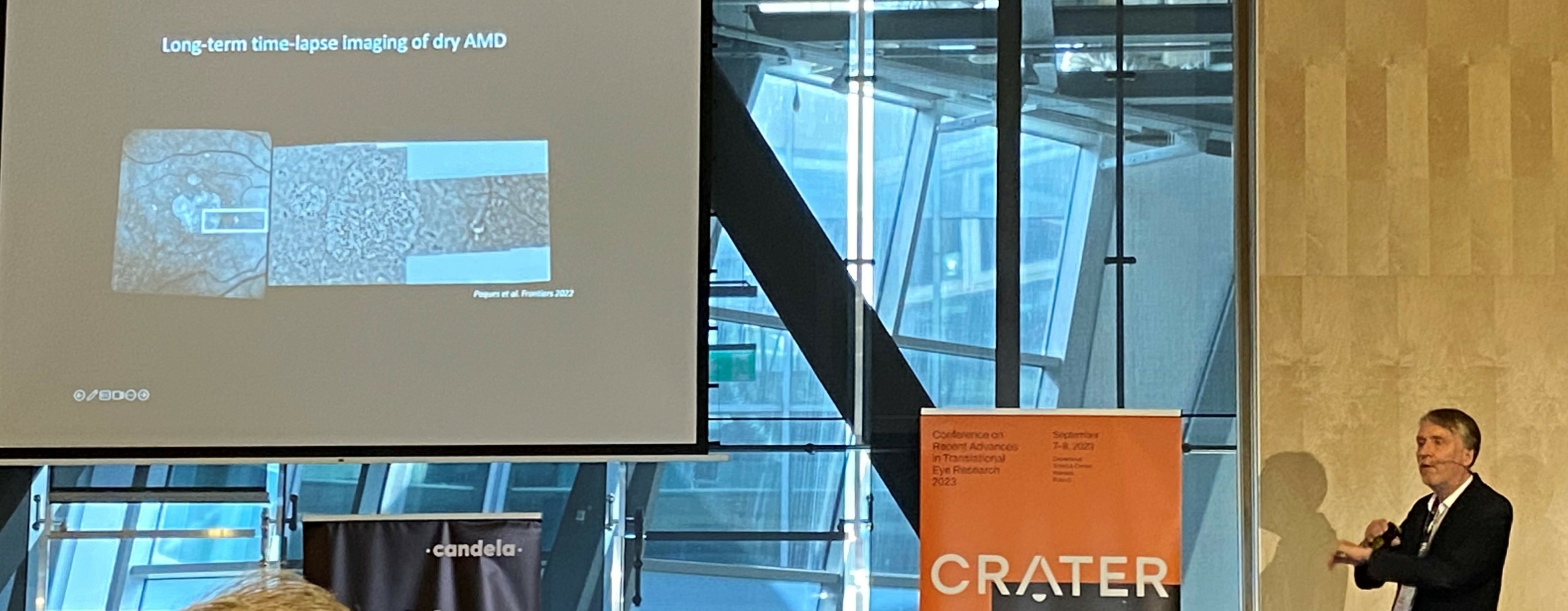
When AMD patients with geographic atrophy are examined with rtx1 Adaptive Optics retinal camera, the border of GA lesions becomes visible at the microscopic scale. It also reveals small hyperpigmented spots, that were shown to redistribute spatially over time [1, 2, 3].
Today at CRATER, Prof. Paques discussed the relationship between this redistribution of hyperpigmented spots and the enlargement of atrophic lesions. His presentation reviewed multimodal and longitudinal images from AMD patients. OCT, SLO, and AF images were completed by time-lapse sequences of rtx1 AO images, as well as novel transscleral images obtained with the TFI optional module.
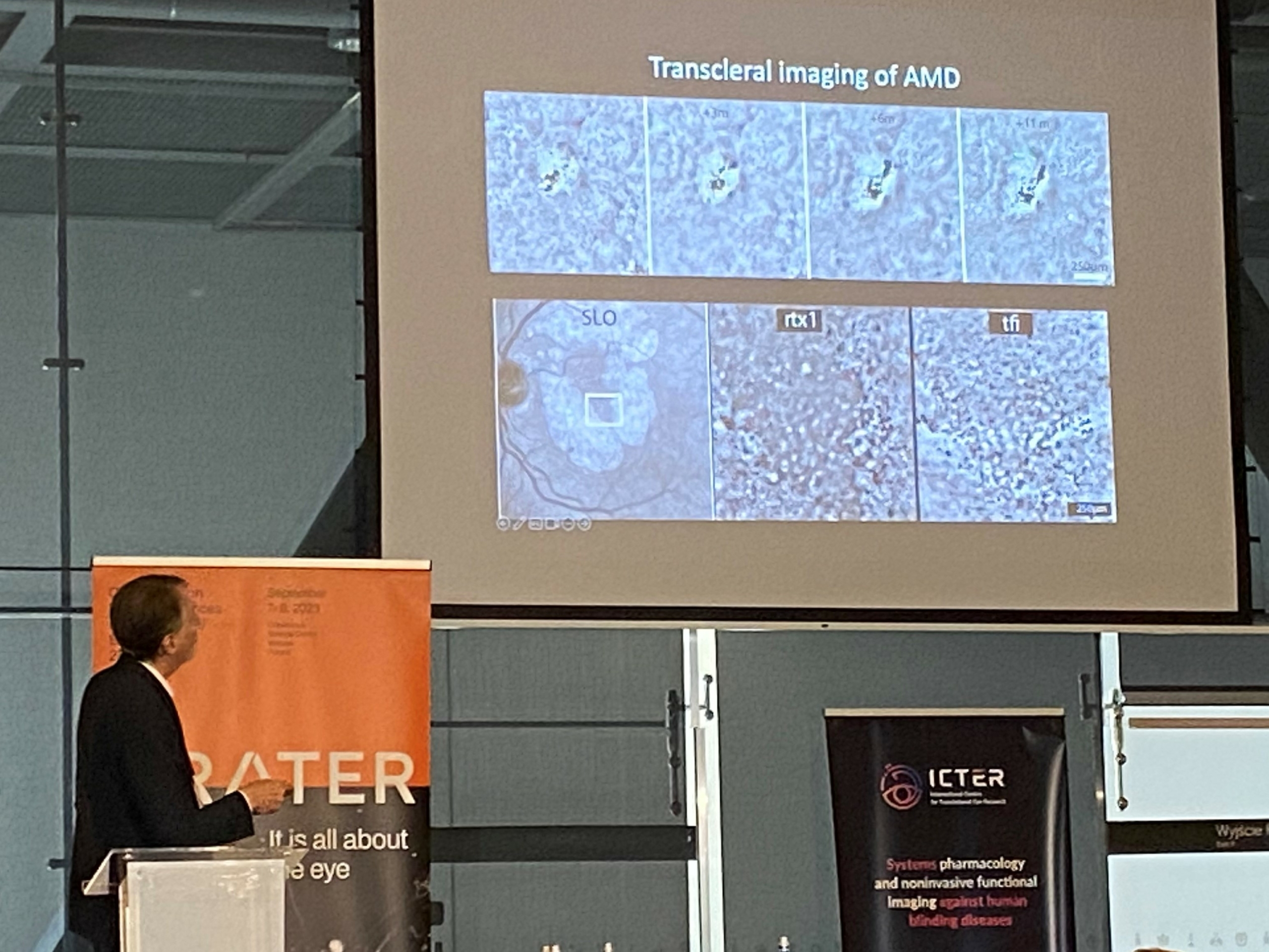
Such data provided information at smaller scales – both dimensional and timewise – than the gold-standard methods, which contributes to a better understanding of cellular dynamics underlying GA progression.
CRATER conference, on 2023/09/07: Documenting the spatial redistribution of hyperpigmented spots during enlargment of geographic atrophy, Michel Paques.
Cited references:
1. Gocho, K., et al. (2013). Adaptive Optics Imaging of Geographic Atrophy. Investigative Ophthalmology & Visual Science, 54(5), 3673 3680.
2. Querques, G., et al. (2016). Adaptive Optics Imaging of Foveal Sparing in Geographic Atrophy Secondary to Age-Related Macular Degeneration. Retina, 36(2), 247 254.
3. Paques, M., et al. (2018). Adaptive Optics Ophthalmoscopy : Application to Age-Related Macular Degeneration and Vascular Diseases. Progress in Retinal and Eye Research, 66, 1 16.
