Therapy assessments in animal models with AO retinal imaging
Two articles recently illustrated the benefits of using adaptive optics (AO) retinal cameras for longitudinal investigations of new therapies in large-animal models, with the advantage of a high translatability to human studies.
Researchers at National Eye Institute (NEI), NIH, demonstrated a laser-induced porcine model for testing therapies for treating retinal degenerations. In their multimodal assessment protocol, the use of a rtx1 AO camera enabled follow-up analyzes of photoreceptor damage.
“AO data further enhances the clinical relevance of this model to develop treatments for human retinal degenerative diseases “
Barone et al. 2023
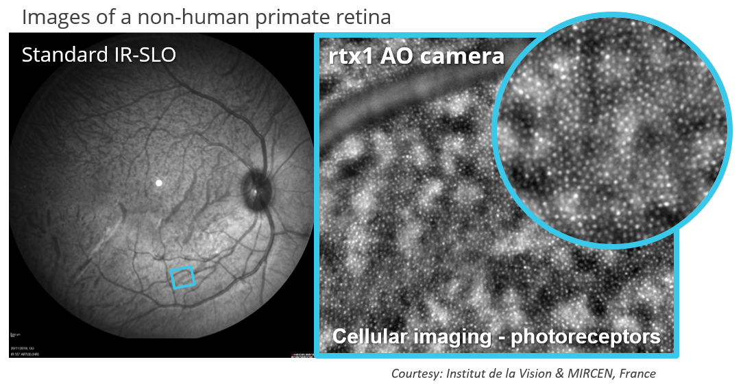
A team at Paris’ Institute of Vision investigated the safety of subretinal injection procedures, which have been increasingly used for delivering retinal therapies. Images acquired with a rtx1 camera allowed them to quantify alterations in photoreceptors and measure their recovery time after the injection.
“We further demonstrate that the AO flood-illumination ophthalmoscopy provides evidence of photoreceptor structural alterations despite no functional change after a subretinal detachment “
Dentel et al. 2023
Article references:
- Barone, F. et al. A versatile laser-induced porcine model of outer retinal and choroidal degeneration for preclinical testing. JCI Insight 8, e157654 (2023). https://doi.org/10.1172/jci.insight.157654
- Dentel, A. et al. Adaptive Optics Flood Illumination Ophthalmoscopy in Nonhuman Primates: Findings in Normal and Short-term Induced Detached Retinae. Ophthalmology Science 3, 100316 (2023). https://doi.org/10.1016/j.xops.2023.100316