2-second acquisitions
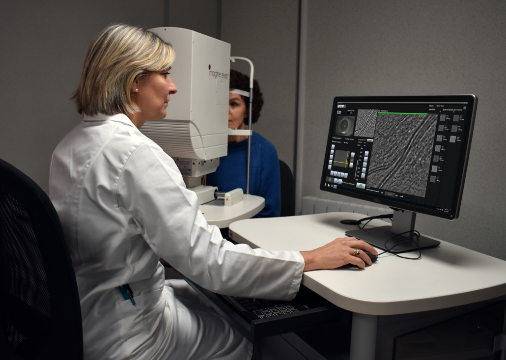
No compromise on patient comfort
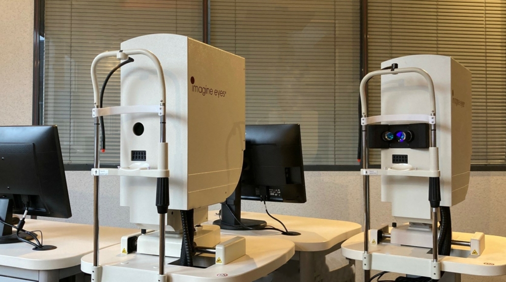
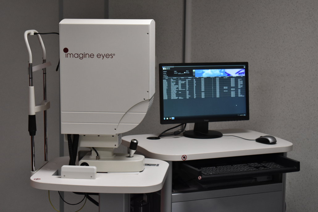
Adaptive optics retinal imaging device with regulatory approval in several countries
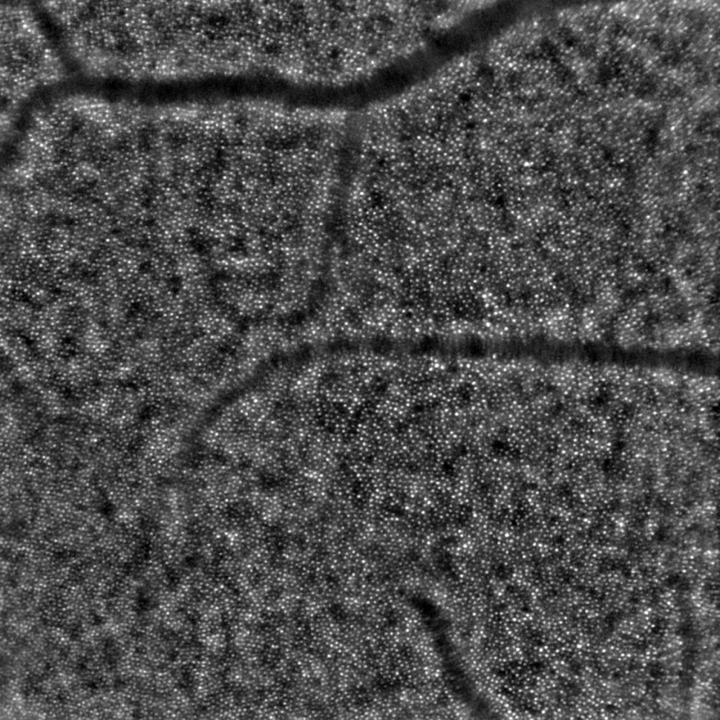
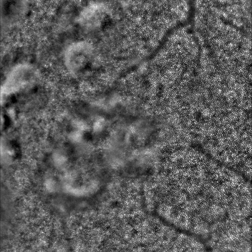
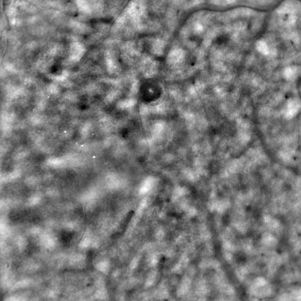
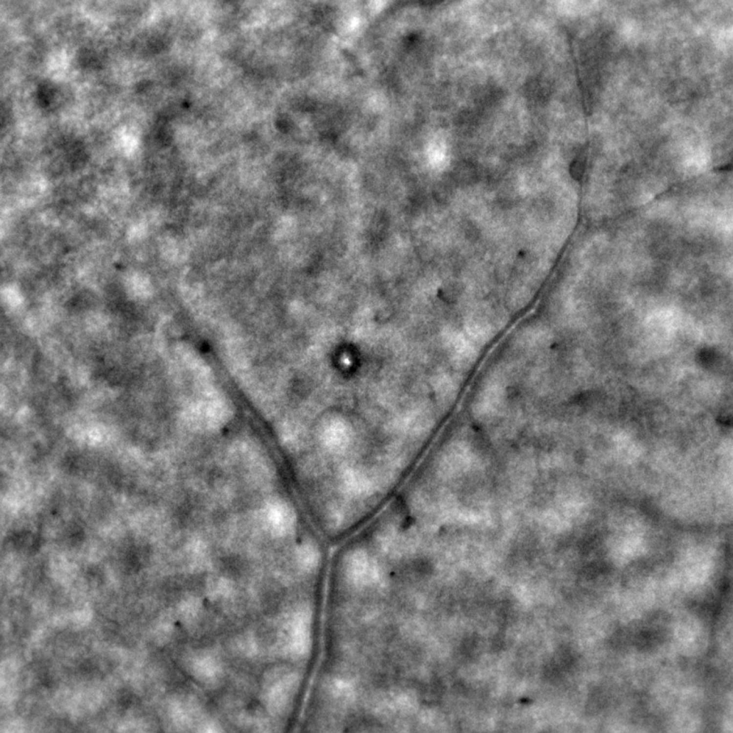
When using the rtx1 Adaptive Optics Retinal Camera, you examine the retina at a scale where individual cells are visible. It reveals parafoveal cone photoreceptors as well as other microscopic retinal structures that cannot be seen with conventional techniques, thanks to a 10 times better resolution.
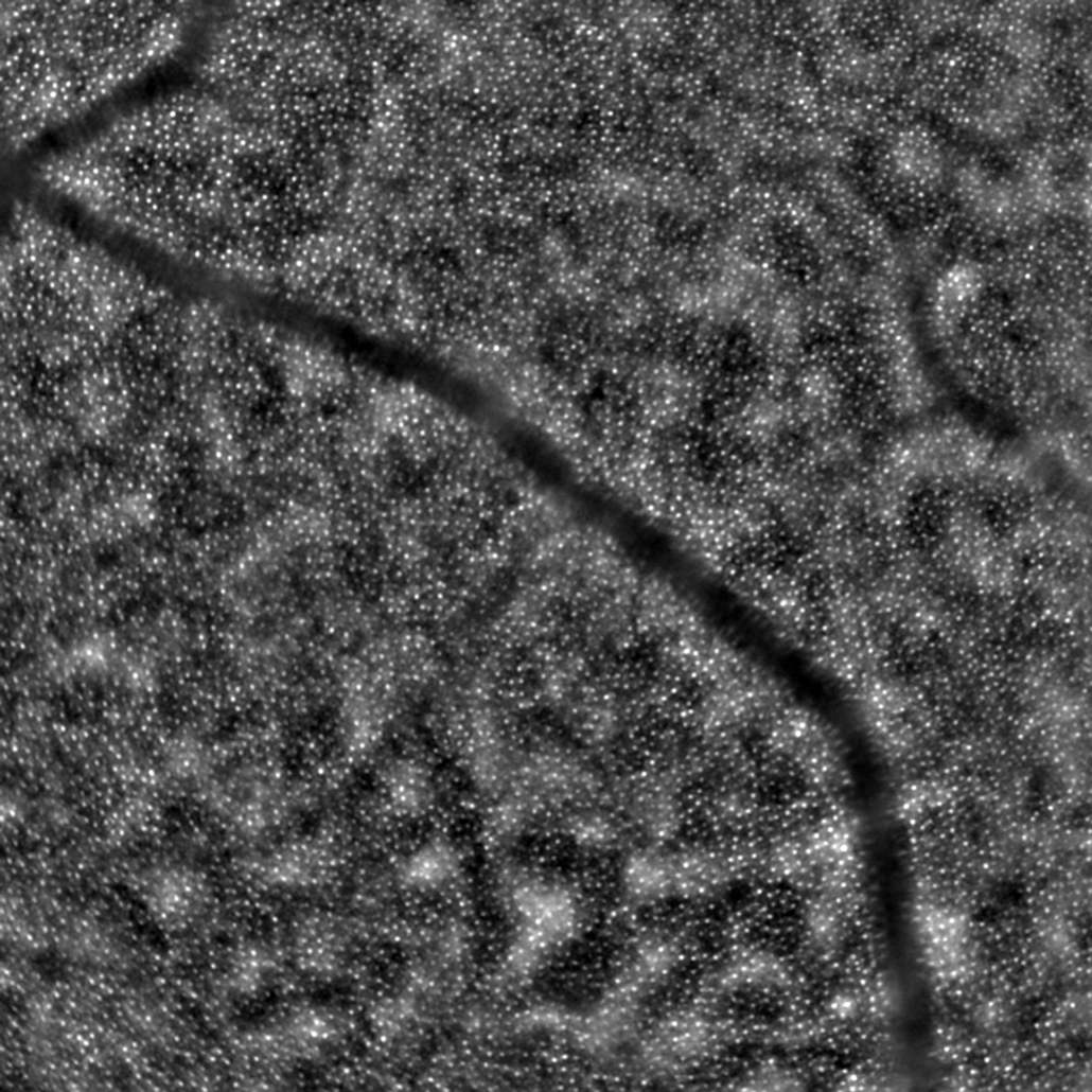
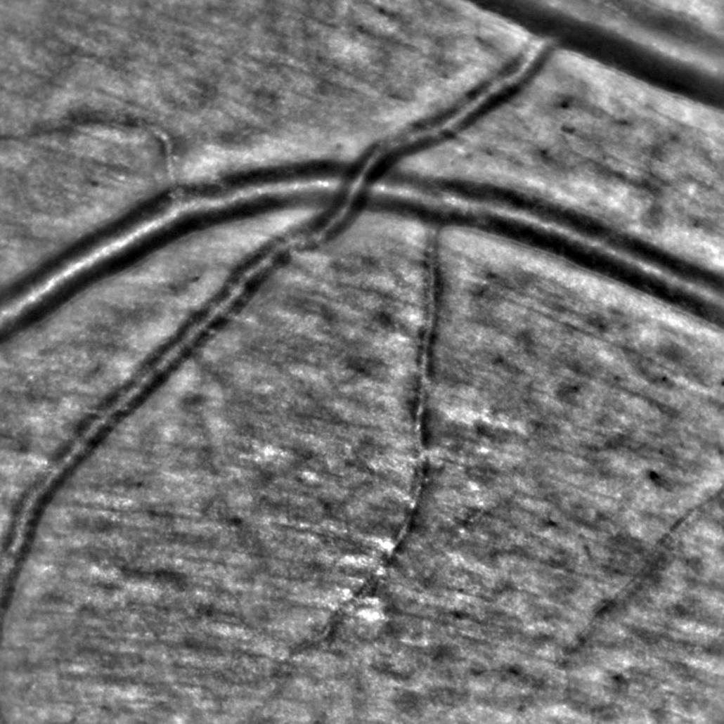
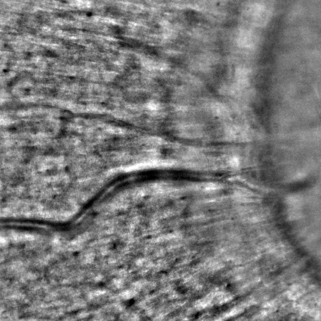
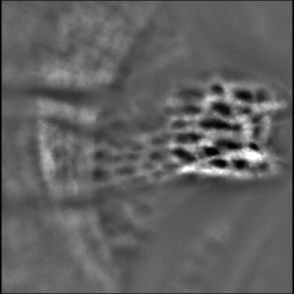
The rtx1 enables visualizing the microscopic walls of retinal arterioles non-invasively, thanks to adaptive optics imaging. Focal narrowing, perivascular sheathing, micro-hemorrhages and micro-aneurisms are also visible without using contrast agents.
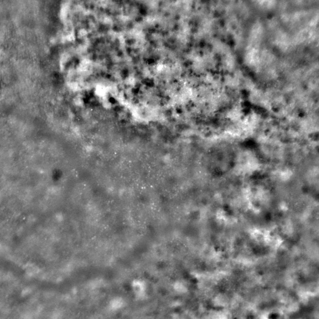
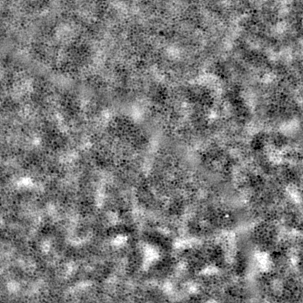
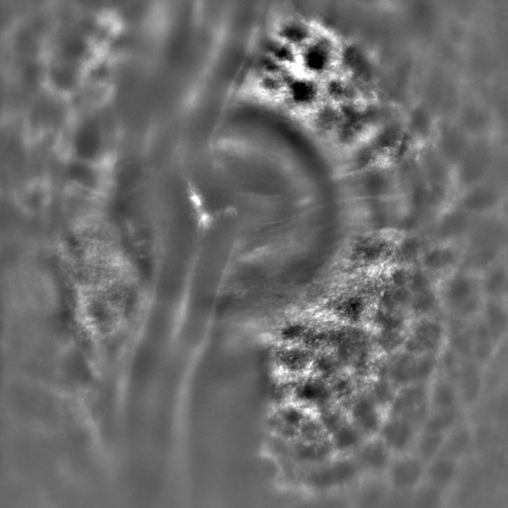
The TFI add-on module extends the imaging capability of rtx1 for investigating retinal anatomy, pathology and therapy. Using transscleral illumination, rtx1-TFI enables visualizations of otherwise invisible RPE cells and pigment redistribution in retinal degenerations.
AO examinations wiht rtx1-TFI are efficient, as a single acquisition delivers co-registered images of the cone mosaic and retinal pigment epithelium.
No compromise on patient comfort
Designed for multicenter trials
Micrometric progressions revealed
Fast annotations and analyzes
Automated montage with DualAlign®
Extensively validated technology
The rtx1 stands out as the most widely-used adaptive optics retina imaging device throughout the world. Cleared by regulatory authorities in several countries, and adopted by 90 clinical centers, it has become the reference device for cellular-level retinal assessments in clinical research. More than 250 publications illustrate the extensive clinical knowledge and findings that users of the rtx1 AO camera have gained in a large variety of diseases that impact the retina.
Designed in collaboration with clinicians, the rtx1 enables comfortable and quick examination for high patient throughput.
The Protocols functionality guides users through every step of virtually any imaging procedure. It enables accelerating and standardizing AO examinations in multicenter studies.


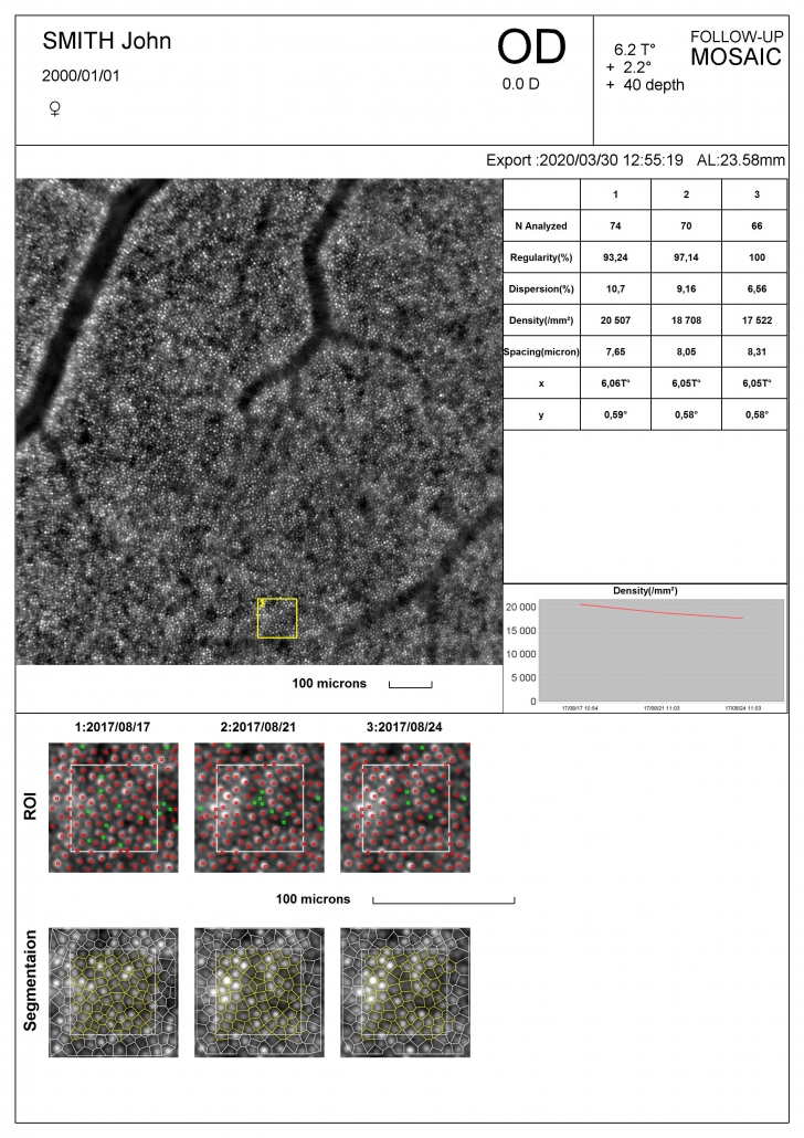
By design, the rtx1 delivers images that are free from motion distortion. Building on this advantage, the software makes it easy to capture multiple regions of interest through different visits, and automatically registers follow-up images. This allows tracking minute changes in groups of cells, vessels, or lesions over time.
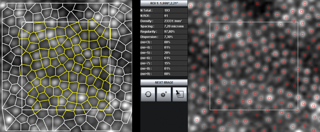
With AOdetect application, rtx1 delivers an array of candidate biomarkers for assessing the distribution of parafoveal cone cells, as well as RPE cells on TFI images :
On follow-up images, AOdetect enables analyzing the exact same region of interest, for monitoring a given group of cells over time.
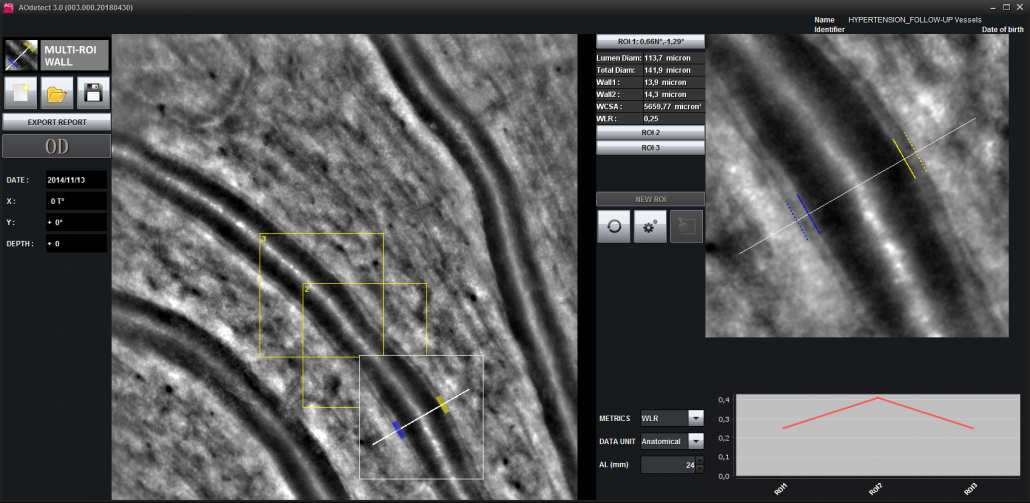
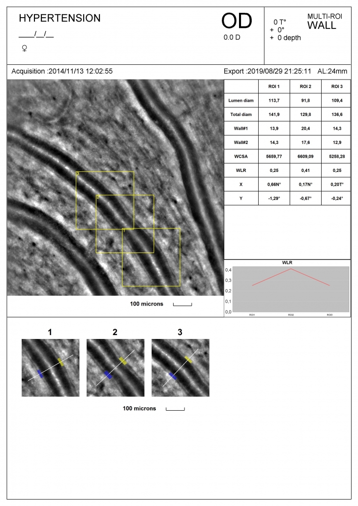
AOdetect application also provides supervised image segmentation for analyzing the wall morphology of blood vessels. With this option, rtx1 delivers non-invasive vascular structure metrics with micrometer precision:
The exact same vascular section is assessed on follow-up images with a only a few clicks.
With rtx1, Flicker stimulation combined with adaptive optics retinal imaging enables micron-precision assessments of neurovascular response in health conditions and diseases that affect small blood vessels.
The rtx1 software provides guided procedures for exploring alterations of retinal microstructure
Gaze-dependent imaging which has enabled visualizations of very small drusen with enhanced contrast 1
Pupil-dependent imaging that allows investigating photoreceptor misalignment in pathologies 2
[1] Rossi et al. Trans. Vis. Sci. Tech. 10, 19 (2021)
[2] Paques et al. Retina 10.1097 (2020).
rtx1 images are compatible with the first-in-class registration algorithms from DualAlign®. With i2k Retina AO™ software, effortlessly assemble multiple AO images into high quality montages.
NEW for clinical research
AObox™ is an AI-based application for analyzing the cone mosaic captured by adaptive optics retinal cameras, co-developed by Erasmus MC and Imagine Eyes. Fully compatible with rtx1 AO cameras, it generates cone density heatmaps and metrics – even directly from multi-image montages.
")


")
")


| Imaging type | En face reflectance imaging |
| Detection type | Low-noise CCD camera |
| Illumination | Near infrared LED, 850nm |
| Exposure time | < 10 ms |
| Imaging field of view 1 | 4° x 4° |
| Fixation stimulation range | H ± 14.5° / V ± 10° |
| Camera pixel pitch on the fundus 1 | 1.1 µm |
| Optical resolution on the fundus 1, 2 | 250 line pairs per millimeter (lppmm) |
| Adaptive optics control | Fully automated, resistant to blinking and movement |
| Depth focussing range 1 | 1600 µm |
| Pupil diameter | ≥ 4 mm |
| Refractive error compensation | -12 to +6 D |
| Total footprint (WxDxH) | 137x53x132-162 cm |
1 Some specifications are dependent on several factors including but not limited to: ocular biometry, pupil diameter, optical defects, ocular media transparency.
2 The system can image line pairs of 2 µm in line width.

rtx1 is a certified medical device of class IIa in the European Union. rtx1 is an approved medical device in Japan, China and South Korea. In the USA, rtx1 has not received FDA clearance; it is an investigational device that requires Institutional Review Board (IRB) oversight. For use by trained eyecare professionals only.
The contents of this webpage may differ from the curent state of approval of the product in your country. Please contact Imagine Eyes or your local representative for more information.
i2k Retina is not part of the rtx1 product, it is for research use only. DualAlign, i2k, i2k Retina AO are trademarks of DualAlign LLC.
AObox is not part of the rtx1 product, it is an application for research use only, developped for rtx1 AO images. AObox is property of Erasmus MC Rotterdam. Its development was supported by Health Holland, Top Sector Life Sciences & Health under grant agreement no. EMCLSH22014 (AO-VISION).
Some retinal images on this page are courtesy of: Quinze-Vingts National Eye Hospital, Paris, France.


