Deciphering AMD: cellular phenotyping & short term progression
On September 20, 2022, we had the pleasure to interview Profs. Michel Paques, Andrew Lotery and Paulo-Eduardo Stanga about the use of adaptive optics (AO) imaging in age-related macular degeneration (AMD). We are grateful to these 3 ophthalmology experts who kindly shared their experience and personal insights. If you missed the live session, click below to watch the 25-minute replay.
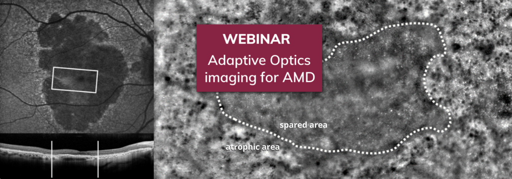
In this webinar, our guests explained how rtx1 AO cameras provide new and complementary information to other retinal imaging techniques, such as fluorescence angiography and high-resolution OCT. According to them, it enables further understanding of AMD mechanisms and it adresses unmet needs in the current context of novel therapeutic developments.
When sharing some example images, the experts described the highly detailed AO phenotypes that they could observe at different stages of AMD. They even showed how time-lapse imaging with rtx1 reveals the dynamics of pigment redistribution, and how it detects geographic atrophy progression over unprecedentedly short timescales.
By pushing forward their investigations, their goal is to identify early biomarkers of AMD progression and of treatement effectiveness, that could be used as surrogate endpoints. Combined with better patient selection, it would enable more targeted and shorter clinical trials for new therapies. AO findings in AMD clould also enable a more personalized clinical follow-up of patients.
About our guests:
Andrew Lotery, MD, FRCOphth, is Professor of Ophthalmology at NHS University Hospital Southampton research interests include investigating the molecular basis of AMD, a condition he develops treatments for by conducting clinical trials. He currently leads an international research project funded by the Wellcome Trust, PINNACLE, that evaluates adaptive optics imaging as a tool to better understand the progression of AMD.
Michel Paques, MD, PhD, is Professor of Ophthalmology and researcher at Quinze-Vingts National Eye Hospital and Vision Institute, in France. His focus is on medical applications of high resolution in vivo retinal imaging in humans. He pioneered the development of cell-specific imaging using SLO, and the application of adaptive optics ophthalmoscopy for a variety of retinal diseases, including geographic atrophy secondary to AMD.
Paulo Eduardo Stanga, MD, PhD, is Director of The Retina Clinic London and Professor of Ophthalmology at the University College London. Since 1993, in addition to treating patients, he has worked in the development and application of new clinical and surgical therapies and technologies, such as AO imaging of photoreceptors and retinal pigment epithelium. His current research activities include, amongst others, clinical studies on gene therapy surgery & anti-complement injections for dry AMD.