New technique detects small drusen in AMD
In a joint study in age-related macular degeneration (AMD), clinical researchers at the University of Pittsburgh, USA, and Quinze-Vingts National Eye Hospital, France, assessed a new method for visualizing drusen using adaptive optics (AO) images. They have called it gaze-dependent AO imaging.
The results obtained in healthy volunteers and patients with AMD were recently published in Translational Vision Science & Technology. In gaze-dependent AO images obtained with rtx1 AO cameras, drusen appeared as annular structures with high contrast and clear delimitation, suitable for quantification.
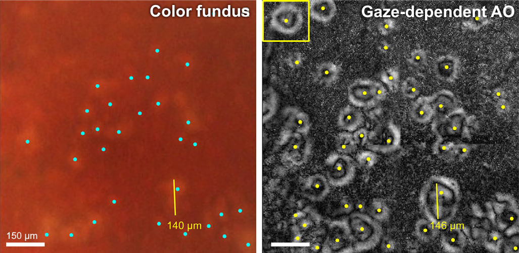
The technique enabled increasing the detection of drusen by up to 250% compared to the gold-standard method using color-fundus images.
“Gaze-dependent imaging may be useful to document the earliest stages of aging of the photoreceptor-RPE complex, particularly the smallest drusen (<25 μm), and to document the dynamics of early drusen progression. “
Article reference: Rossi, E. et al. A New Method for Visualizing Drusen and Their Progression in Flood-Illumination Adaptive Optics Ophthalmoscopy. Trans. Vis. Sci. Tech. 10, 14 (2021). DOI: 10.1167/tvst.10.14.11
