Investigation with rtx1 shows how myopia affects the arrangement of retinal cells
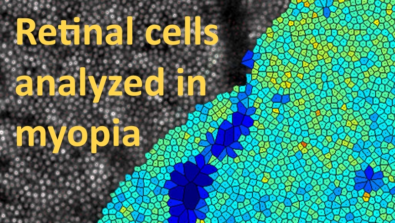
A study published in Ophthalmic and Physiological Optics used the rtx1 Adaptive Optics Retinal Camera to assess the distribution of cone photoreceptor cells in myopic subjects.
The authors, Kelly Woog & Richard Legras at the University of Orsay, France, reported that retinas in myopic eyes had significantly lower cone cell density when compared with normal emmetropic eyes. According to the results, the difference was in the order of 10%. The reduction in cell density is believed to be caused by stretching of the retinal tissue, which is associated with the elongation of the eyeball that usually accompanies the development of myopia.
While reading the paper, a question arises: does the lower-than-normal cone cell density of myopes affect their visual performance?
Although the study did not assess visual performance, the authors provide useful insight. On the one hand, when the eyeball becomes longer, the retina stretches and the distance between neighboring cells increases: these changes tend to alter the eye’s ability to discriminate small details. On the other hand, the eyeball elongation also augments the optical magnification of the eye, which tends to enhance the vision of small details. In other words, there are two effects that seem to compensate each other. This may explain how myopic eyes -once their refractive error is corrected with lenses – can achieve similar performance as normal emmetropic eyes.
These new results illustrate how adaptive optics retinal imaging can provide deeper insight not only into retinal pathologies, but also in other widespread conditions like myopia.
To learn more about the rtx1 Adaptive Optics Retinal Camera : click here