Inherited retinal diseases: advances in imaging to accelerate progress in therapy
22.09.2021
Highlights of the Euretina 2021 meeting
Inherited retinal diseases (IRDs) are a major cause of blindness in children and working-age adults. Recent advances in genetics and molecular biology have fostered the development of several treatments for IRDs, with the initiation of multiple clinical trials and the approval of the first gene therapy.
In this promising context, retinal imaging techniques are essential to acquiring knowledge in the phenotypes and natural progression of IRDs. Retinal imaging also has a major role to play in the stratification of trial volunteers, the identification of positive responders, the control of treatment delivery, the evaluation of effectiveness, and the detection of side effects.
Based on optical coherence tomography (OCT), quantifications of residual photoreceptor cells have been proposed as inclusion criteria and outcome measurements. However, with this technique, it takes 12 to 24 months to detect modifications caused by IRD progression. This is due to the limited image resolution of current OCT technology, which does not allow seeing individual retinal cells.
Adaptive optics (AO) technology has overcome this limitation. The rtx1 AO camera commercialized by Imagine Eyes has enabled clinicians to track diseases at the cellular level in the retina. At the 2021 Euretina meeting, several presentations have illustrated the readiness and benefits of AO cameras for investigations in IRD therapies:
- A team at the University of Tuebingen, Germany, used rtx1 to monitor the rescue of cone photoreceptors after gene therapy with voretigene neparvovec (Luxturna®) in Leber congenital amaurosis. The authors stated that the images demonstrated:
“a remarkably fast restoration of the central photoreceptors within only 5 weeks after treatment”
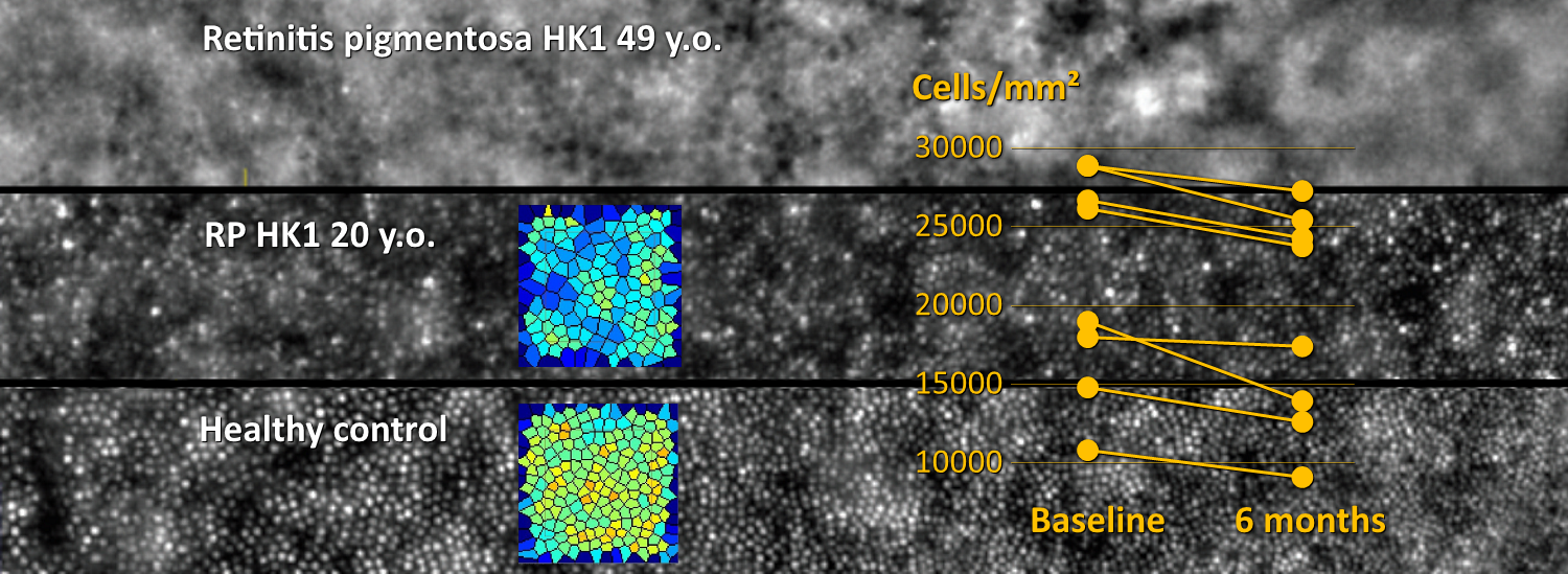
- In retinitis pigmentosa caused by a mutation in the HK1 gene, ophthalmologists at the Nippon Medical School, Japan, measured a 40% loss in cone cells with rtx1, while no damage was quantifiable in OCT images. The authors explained:
“the disease progression is slow, and symptoms begin in the late 40s […] AO revealed that the cone photoreceptor densities were significantly reduced in the parafoveal area at the age of 20 years”
- In a cohort of patients affected with retinitis pigmentosa, clinical researchers at the Lions Eye Institute, Australia, measured an 11% drop in cone density with rtx1 over a period of 6 months, while OCT detected no progression over the same period of time. The authors concluded:
“parafoveal cone density should be considered as an endpoint in future natural history studies and clinical trials in RP”
These brilliant reports demonstrated short-term assessments of progression and regression of cellular damage caused by IRDs, beyond the possibilities of current OCT devices. The results advocate the use of AO retinal cameras in clinical research studies, natural history studies, concept studies, as well as therapeutic trials. Cellular resolution will benefit most investigations in IRDs.
At Imagine Eyes, our team helps implement AO imaging and optimize its use in each particular context. We are keen to provide continuous support to investigators, trial sponsors and CROs in their fight against IRDs.

