Clinical evidence: RPE cells imaged with rtx1-TFI
In a study published in Diagnostics, clinical researchers at Quinze-Vingts National Eye Hospital, INSERM and University of Pittsburgh School of Medicine investigated the retinal pigmented epthelium with rtx1-TFI, an adaptive optics retinal camera with transscleral illumination.
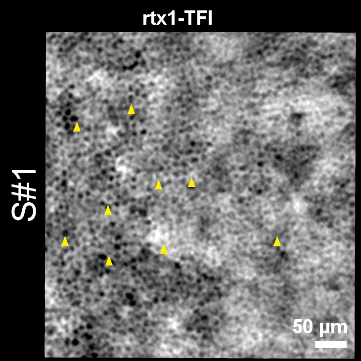
They examined the same eyes with a method capable of revealing RPE cells – near-infrared autofluorescence AOSLO – and with the transscleral imaging modality of rtx1.
For the first time, the authors report cell-to-cell comparisons between TFI and NIRAF images. It provides clinical confirmation that rtx1-TFI enables assessing RPE cells.
“If we compare the time needed for capturing the same retinal area, rtx1-TFI is 35 times faster, which makes this system adapted to a clinical environment.”
To learn more, see also the interview with ophthalmologists Prof. Michel Paques, Prof. Kiyoko Gocho and Dr. Ysé Borella, who kindly shared their insight on how rtx1-TFI enables investigating pigment migration in dry AMD.
TFI is an optional add-on for the rtx1 AO retinal camera. It is available as an upgrade and is compatible with most existing rtx1 units.
Article reference: Gofas-Salas, E., Lee, D. M. W., Rondeau, C., Grieve, K., Rossi, E. A., Paques, M., & Gocho, K. (2024). Comparison between Two Adaptive Optics Methods for Imaging of Individual Retinal Pigmented Epithelial Cells. Diagnostics 14(7), 768. https://doi.org/10.3390/diagnostics14070768.