Regenerative retinal therapy evaluated with adaptive optics imaging
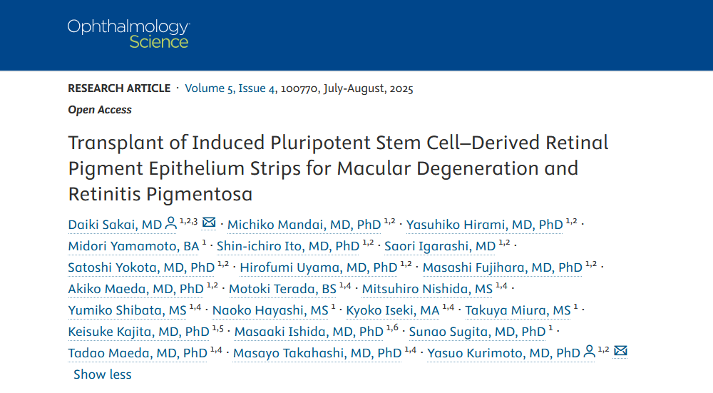
A recent study published in Ophthalmology Science used adaptive optics (AO) retinal imaging in the first human investigation of a regenerative therapy for macular degeneration. The new procedure, developed at Kobe City Eye Hospital in Japan, involved transplanting strips of retinal pigment epithelium (RPE) cells derived from allogeneic induced pluripotent stem cells (iPSCs).
Standard functional tests, such as microperimetry, were inconclusive in this investigation due to the patients’ extremely low baseline retinal sensitivity. However, AO imaging with the rtx1 camera made it possible to visualize the transplanted cells in all participants. Over the 52-week follow-up period, high-resolution AO images revealed a significant expansion of the grafted cell area, providing a structural indicator of therapeutic integration that would otherwise be difficult to assess.
These results underscore the importance of AO imaging for monitoring the outcomes of retinal therapies, particularly when functional measures alone are insufficient.
Read the full article
Learn more about the AO retinal camera used in the study
Article reference
Sakai, D., Mandai, M., Hirami, Y., Yamamoto, M., Ito, S.-I., Igarashi, S., Yokota, S., Uyama, H., Fujihara, M., Maeda, A., Terada, M., Nishida, M., Shibata, Y., Hayashi, N., Iseki, K., Miura, T., Kajita, K., Ishida, M., Sugita, S., … Kurimoto, Y. (2025). Transplant of Induced Pluripotent Stem Cell-Derived Retinal Pigment Epithelium Strips for Macular Degeneration and Retinitis Pigmentosa. Ophthalmology Science, 2025. https://doi.org/10.1016/j.xops.2025.100770
