Natural history study identifies a six-month progression biomarker in retinitis pigmentosa
In a recent publication in Scientific Reports, ophthalmologists from Kobe City Eye Hospital in Japan reported findings from the KEYS study. KEYS is a prospective natural history study that follows patients with retinitis pigmentosa (RP) caused by mutations in the EYS gene.
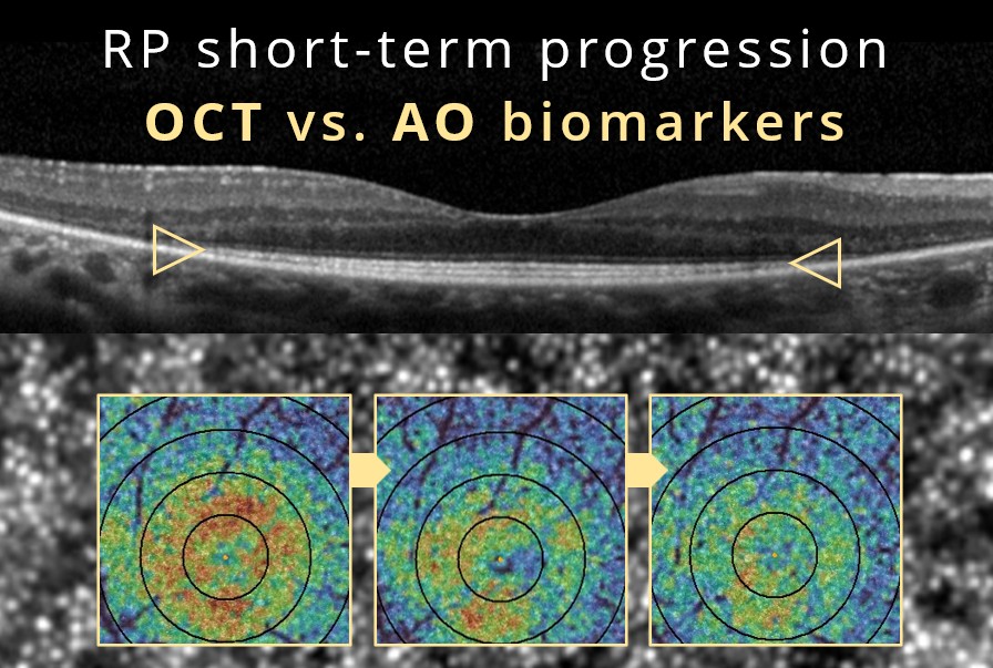
Over the course of two years, the researchers compared structural and functional measures in 27 patients. Visual field testing showed no significant change at any point during the study. Changes in visual acuity only reached statistical significance after 18 months. OCT-based ellipsoid zone measurements took a full 24 months to show a significant reduction. In contrast, cone photoreceptor density, as measured with an adaptive optics (AO) retinal camera, showed a significant decline in every assessed region at each six-month follow-up throughout the two-year study.
“The findings of our study suggest that cone density changes may be detected earlier than functional measures in EYS RP. While reductions in ellipsoid zone width were significant at 24 months, cone density showed significant changes much earlier, reinforcing its potential as an early structural indicator of disease progression”
These findings echo and expand upon the earlier work of ophthalmologists at the Lions Eye Institute in Perth, Australia. Using an AO retinal camera, the researchers found significant cone loss within six months in patients with rod-cone dystrophy, even though OCT showed stable ellipsoid zone integrity.
Together, these studies demonstrate that AO imaging can measure disease activity at the cellular level long before it becomes visible on conventional OCT scans. This capability is directly relevant to natural history studies, proof-of-concept work, and therapeutic trials in inherited retinal disease. A fast, sensitive endpoint can meaningfully shorten the time needed to demonstrate a treatment effect.
If you are evaluating AO biomarkers for your own research, contact Imagine Eyes. We specialize in integrating AO imaging into clinical protocols and optimizing its use for specific diseases and endpoints.
References
Hiraoka, M. et al. Two-year prospective natural history study of EYS-associated retinitis pigmentosa using adaptive optics: the KEYS study. Sci Rep 16, 25938 (2026). https://doi.org/10.1038/s41598-026-56837-3
Roshandel, D. et al. Short-Term Parafoveal Cone Loss Despite Preserved Ellipsoid Zone in Rod Cone Dystrophy. Trans. Vis. Sci. Tech. 10, 11 (2021). https://doi.org/10.1167/tvst.10.14.11
