Photoreceptor cells quantified in RP with and without CME
Recently published in PLOS ONE, a clinical study at the Kobe Eye Center reports quantitative assessment of cone photoreceptor cells in retinitis pigmentosa patients with and without cystoid macular edema (CME) using a rtx1 AO camera. Parafoveal cones were analyzed in locations where OCT did not show CME.
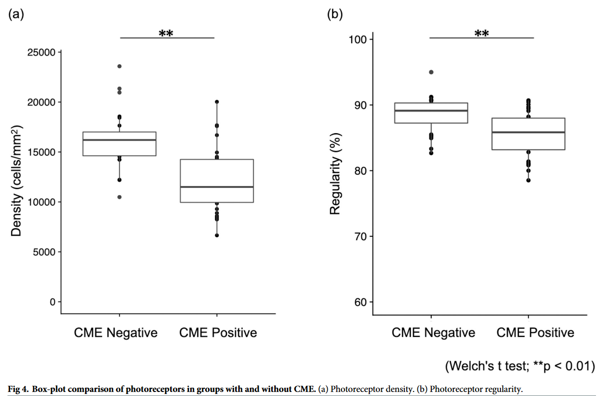
“Although the length of the EZ/IZ, which reflects the progression of RP, did not significantly differ between the groups, a history of CME was associated with photoreceptor loss.”
Article reference: Kitahata, S., Gocho, K., Motozawa, N., Yokota, S., Yamamoto, M., Maeda, A., Hirami, Y., Kurimoto, Y., Kadonosono, K., & Takahashi, M. (2024). Evaluation of Photoreceptor Features in Retinitis Pigmentosa with Cystoid Macular Edema by Using an Adaptive Optics Fundus Camera. PLOS ONE, 14. https://doi.org/10.1371/journal.pone.0296493.