Retinal microvascular imaging beyond OCTA
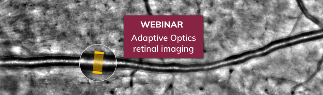
OCTA is a great tool to look at retinal perfusion. Adaptive optics (AO) retinal imaging adds another dimension: it resolves the retinal microvascular structure, unveiling arteriolar lumen and wall , so that alterations can be measured with micrometer precision.
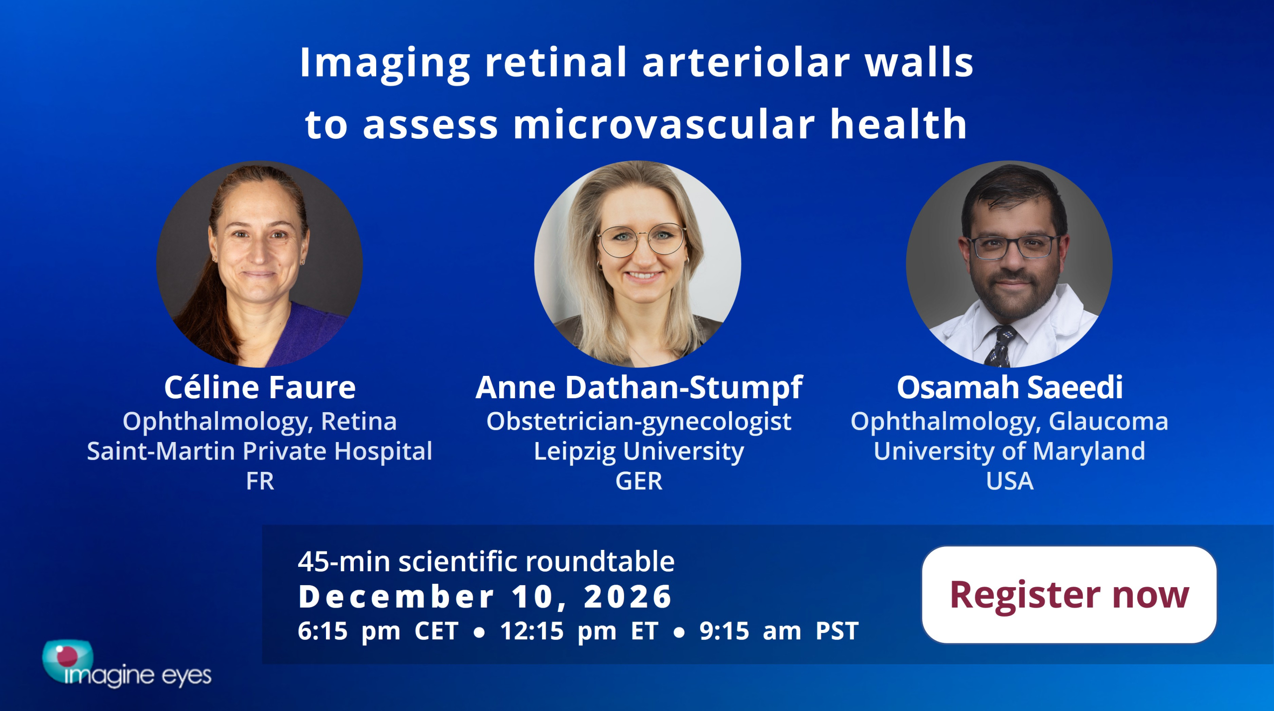
What does this make possible? Three clinicians who use the RTX1 AO retinal camera in their research will discuss applications in a roundtable, followed by an interactive Q&A where you can bring your own questions.
📅 Thursday, December 10 · 6:15 pm CET / 12:15 pm ET · 45 minutes · Live on Zoom
Why attend
Microvascular alterations matter far beyond the retina. Whether you work in ophthalmology, internal medicine, but also cardiology, nephrology, or pregnancy-related research, this session will show what micrometer-scale vascular imaging could bring to your studies.
Format
- Roundtable discussion with 3 panelists
- Interactive Q&A session
Panelists
- Céline Faure, MD PhD, ophthalmologist, Saint Martin Private Hospital, Caen, France
- Osamah Saeedi, MD MS, ophthalmologist (glaucoma), University of Maryland School of Medicine, Baltimore, USA
- Anne Dathan-Stumpf, MD, obstetrician-gynecologist, Leipzig University Hospital, Germany
Please register to join the live session
Want to know more?
Wondering how rtx1 could fit into your research?
Looking for the published evidence on AO microvascular imaging?
